How do I identify a suspicious lesion?
It is important to know your skin and what’s normal for you. This can be accomplished by monthly self-skin examinations.
All you need are your eyes and a mirror.
Checking over your body regularly for any new growths and changes to preexisting moles is important to help identify suspicious lesions. Address any new, changing, or rapidly growing lesions with your doctor or dermatologist.
Monthly self-examinations combined with routine dermatologic evaluations can greatly improve the likelihood of catching skin cancer early and treating it effectively and efficiently.
When completing your self-examination, here is what to look for:
- Changes in the size, shape, or color of a mole or growth.
- A lesion that is rough, oozing, bleeding, or scaly.
- A sore lesion that will not heal.
- Pain, itching, or tenderness to a lesion
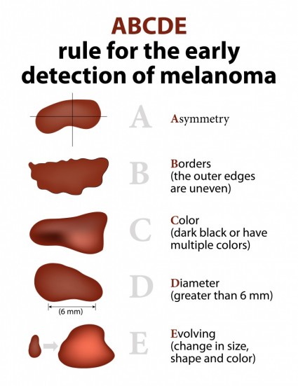
The ABCDEs of identifying a melanoma:

These guidelines can help you identify suspicious areas to discuss with your doctor.
- Asymmetry: the two halves of a lesion do not match
- Borders: the edges of a lesion are blurred, uneven, or irregular
- Color: the color throughout the lesion is not consistent. It may contain shades of black, brown, red or white
- Diameter: a lesion that is greater than 6 millimeters (about ¼ inch) across its diameter
- Evolving: changes in the mole’s size, shape, and/or color
Click here to watch a video about performing a self skin-examination.
If your dermatologist suspects you have skin cancer, a small biopsy can be performed to obtain a diagnosis.
Types of skin cancers:


